Using Chlamydomonas reinhardtii as a model organism, we wished to
investigate how cilia movement turns into propulstion, how cilia coordinate with
each other hydrodynamically, and in general, develop techniques to manipulate cilia.
This work was done by Andrew Fung and Hsiang-wei Lu during the Bioengineering Bootcamp
of September, 2007.
The bulk of the three days devoted to the project involved obtaining a high percentage
of reactivated cilia after purification, and the attachment of polystyrene microspheres to
the cilia as handles to use with optical tweezers.
Experimental Methods
The protocol for cilia purification was developed by a cohort of scientists in the
mid-70's by Borisy, Gibbons, Mitchell, and Witman, among many others. We opted for a
relatively simple method, that of using dibucaine plus shear forces to strip the
densely-grown Chlamydomonas (grown in TAP medium) of their cilia, then
stripping the cilia of their membranes to facilitate bead attachment.
In order to get beads to attach to the cilia, we chose two pathways: 1) nonspecifically
absorbing anti-alpha-tubulin antibodies onto the surface of the 1 micron diameter spheres, and
2) incubating antibody with protein-A coated spheres (protein A being a streptococcus protein
that binds the Fc region of antibodies).
Results
Our method for cilia purification worked well, but there was only a small percentage of reactivated
cilia. If we watched long enough, it was apparent that some were undergoing non-Brownian motion, but
they were incredibly slow, and the percentage was low. Below is a movie of cilia undergoing Brownian motion, and
when ATP is added, we can see one cilia executing stereotypical motion.
Brownian motion of free cilia
Directional movement of free cilia
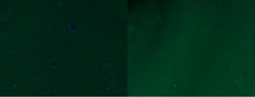
Stripping off the membrane worked well. We stained the cilia with FM464 dye before and after demembranation.
See Figure below. It's an HSV image of a fluorescence image overlaying a brighfield image
in HSV color space. One can see that there is a brighter fluorescence signal in the non-demembranated image (left, in blue).
The long structures are cilia.
For some reason, the bead chemistry did not work that well.
Interactions between a green (532 nm) optical trap and the cilia resulted in a curious observation:
that the cilia will orient along its long axis into the trap. Thus provides an alternate way to handle
the cilia than through bead attachment.