Keratocytes
are the first line of defense when a Cyprinus' (fish/carp)
outer scaled region is damaged. They essentially cover the outside of the fish;
upon injury their connected network is broken. This break in
the network is also accompanied by a small voltage, setup by
the Keratocytes. They then follow the potential gradient to find
the wound
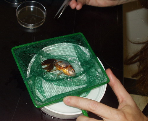
site. Julie Theriot and Eric Kalvins extracted these cells from
a living goldfish (Galvin).
Galvin
was very distressed...but was overall unharmed.
Their
hypothesis was that the cells would exhibit isotropic motion
when there was no potential gradient; but in the presence of
an external potential the internal actin network would align
with the field and polarize the Keratocytes' movement.
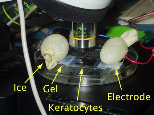
After
the cells were extracted, Julie and Eric designed and built
a small, microscope mounted, galavnotaxis chamber (not bad
for
two
days). Pictured below is their third prototype.
A small liquid viewing chamber was flanked by two electrodes
connected
to a
programmable power source. The heat generated by the cell was
removed with a very sophisticated heat transfer system...two
small bags of ice.
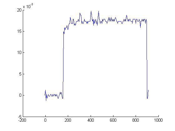
Thus
far, their hypothesis is certainly qualitatively correct. After
some voltage calibration, they applied the following pulse to
the cell, and collected video of the Keratocyte's corresponding
motion. Notice the Keratocytes are at first moving rather randomly,
but once the field is turned on they all proceed downwards.
Voltage
across the cell as a function of time
Corresponding
movie of Keratocyte motility/galvanotaxis.