Optical
tweezers are one the key tools biophysicists are using to unravel
the energetics and kinetic pathways of single, biologically
active molecules and proteins. This method has been used to study
DNA mechanics, viral packaging mechanics and kinetics, RNA and
DNA
polymerization
and a plethora of other topics. The short time the students have
does not enable them to tackle a full, novel project using optical
tweezers, especially considering most students have not worked
with them before this course.
The
aim of this project is three-fold: students become familiar with
the necessary optical techniques and equipment to build the setup
from scratch, they learn the theory necessary to understand why
the particle is trapped and how thermal fluctuations will affect
the particle, finally they write their own software
to analyze the images and determine the 'stiffness' of the trap.
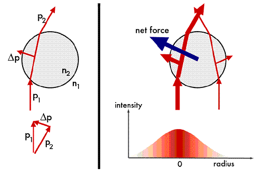
Ray
optics gives us the simplest explanation as to why the particle
remains in the trap despite random thermal fluctuations. The
basic principle is that the light's momentum is conserved through
the scattering process, or in other words, the total momentum
is the same before the light sees the particle as after. The
particle must have a different index of refraction from the surrounding
medium in order to scatter the light and induce a momentum change.
The trap itself is the source of a large optical gradient, which
provides the directionality of the force - always towards the
trap's center.
Diagram
showing net momentum change during light scattering - the intensity
gradient pulls the bead to the trap's center.
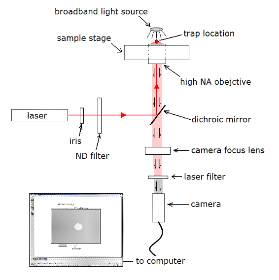
The
students start with a simple diode laser (~30mW @ 632nm)
and must convert this rather 'dirty' beam into a well colimated
laser. The laser is then steered by a series of mirrors to
a dichroic mirror whose reflectance is wavelength dependent.
The laser is reflected by the dichroic through the objective
(100X oil) and the trap forms at the focal point of the objective
(working distance of roughly 250um). A bright field light
source is on the other side of the objective allowing us
to image the particle. The light 'pollution' from the laser
is removed by a series of red light filters, and the final
filtered image strikes a high-speed CCD camera for data collection.
The total strength of the optical trap is regulated by a
variable neutral density filter in the laser's path. This
allows the students to measure the trap strength as a function
of laser power. Finally, as before, the students must spatially
calibrate the objective.
Diagram
showing the basic components of the optical tweezers setup.
Trapping
a particle is not a trivial task as it requires precise
alignment of the laser into the objective, however once aligned
data collection is fairly easy. Videos of the particle are
taken a few minutes at a time in order to gather enough data
to be statistically significant.
Real-time
video of a trapped particle.
With
data in hand, the students then venture into writing Matlab(c)
code to analyze the particle's movement. The particle is
sitting
in an energy well created by the optical trap that is roughly
Gaussian in shape, and hence approximately harmonic near
the trap's center. Statistical mechanics tells us that all
harmonic degrees of freedom contain the same amount of energy
at equilibrium, hence we know:
where k is
the trap stiffness and kT is the thermal energy
unit. This means that a measure of the RMS movement of the
particle directly tells us the stiffness of the trap. Measuring
particle position requires a fair amount of image processing,
the movies are read in as a series of image files; each file
having a calculable x and y position for
the particle.
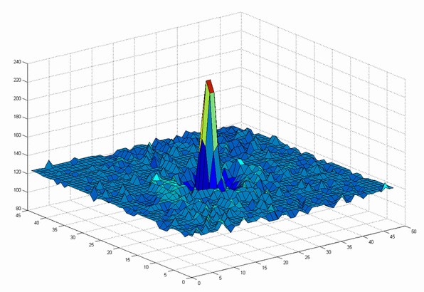
A
single intensity image showing the characteristic peak which
indicates the particle's position.
After
simple intensity adjustments (contrast, thresholding etc)
the particle's x and y position
is tracked using the centroid method, or in other words,
tracking
the
position of the average intensity of the image over time.
A histogram of the particle's position shows the probability
with which the particle was found at any one position.
Histogram of particle's x and y position.
Finally,
this histogram essentially maps out the energy surface felt
by the particle in the trap via the partition function.