10x objective
20x objective
40x objective
100x objective
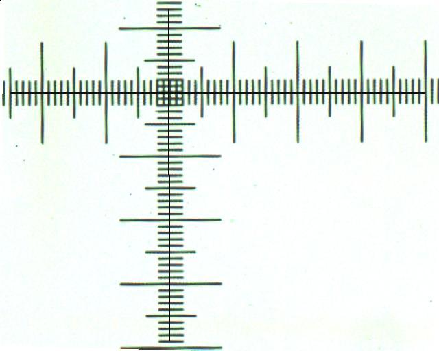
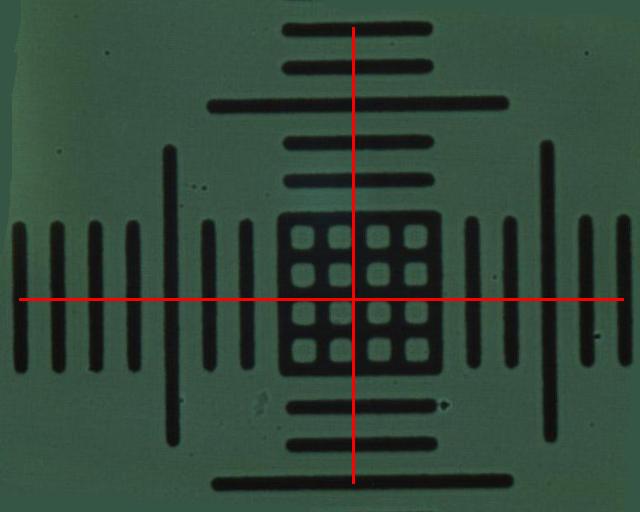
For the last image, we used immersion oil. With these images we acquired the following data with the software ImageJ . Now we can put scale bars on every image.
Magnification |
Length in pixels |
Length in um |
pixels/um |
average |
10x |
605.001 |
650 |
0.931 |
0.932 |
485.001 |
520 |
0.933 |
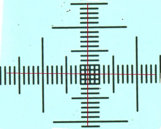
20x |
599.000 |
320 |
1.872 |
1.875 |
488.000 |
260 |
1.877 |
40x |
605.000 |
160 |
3.781 |
3.795 |
457.000 |
120 |
3.808 |
100x |
554.000 |
60 |
9.233 |
9.261 |
555.000 |
60 |
9.250 |
558.000 |
60 |
9.300 |
{top}
Stentor
The origin of its name is very interesting. Stentor is a mythical Greek warrior with an unusually loud voice who died after losing a shouting contest with Hermes. His name has given rise to the adjective "stentorian", meaning loud-voiced. Since some ciliate protozoa had a trumpet-shaped body, they received the same name. Stentor (the protozoa) are a group of filter feeders, that are common in freshwater lakes and streams, usually attached to algae and other detritus.
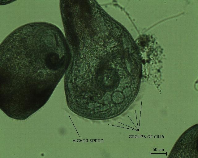
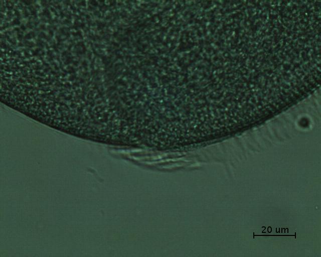
We estimate, using our scale bars, that the length of the Stentor is about 1mm, which makes them one of the largest single-celled organisms.
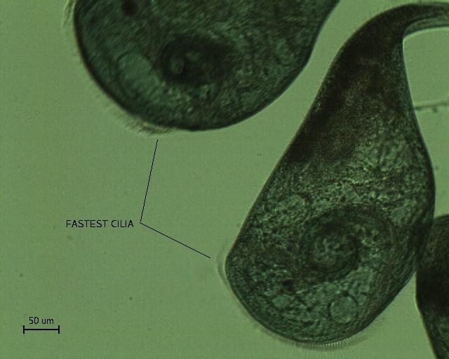
It can be seen that the cilia movement is performed in groups, each moving at a different speed, as it was observed during the snapshots. We also noticed that there was a group of cilia that was always moving faster than the others, as it is indicated in the figure. To estimate the number of cilia that move together, we measured the length of a group (using the image below), which was, on average, around 25um. It was possible to count the cilia, because they were not moving very much:
From four sets of cilia with 25 um, we counted: 7, 7, 8 and 7 cilia. Therefore, there are about 7-8 cilia per moving group of cilia.
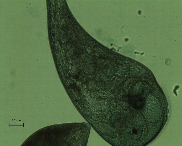
The following images show a closer view on the cilia.
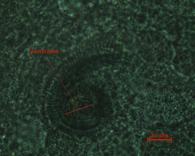
So it is possible to estimate that the cilia are about 10um long and the peristome is about 20um in diameter. The peristome is a region around the cytostome ("mouth" of a protozoan), which receives the food. The food particles get there because the Stentor, using their cilia surrounding the oral surface, creates a vortex.
{top}
E. Coli
Escherichia coli (usually abbreviated to E. coli) is one of the main species of bacteria that live in the lower intestines of warm-blooded animals (including birds and mammals) and are necessary for the proper digestion of food. Its presence in groundwater is a common indicator of fecal contamination. The name comes from its discoverer, Theodor Escherich. It belongs among the Enterobacteriaceae, and is commonly used as a model organism for bacteria in general.
E. Coli cells are rod shape prokaryotic cells with typical diameter of 0.8 um and length 2 um. First we observe E Coli cells under 40x objective and compare the phase contrast image with bright field image.
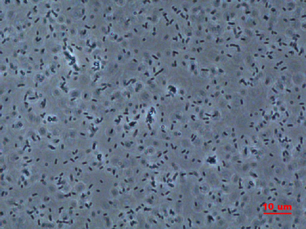
| This is a phase contrast image. The microscope has a phase plate which filters out the original image components. The resultant image is the samples effect on light. |
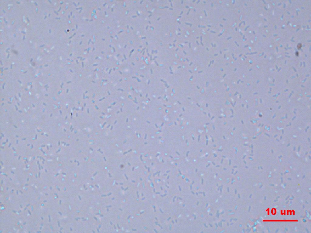
| This is a bright field image. Compared with phase contrast image on the left, the fine features of the sample are not shown as clear. |
As an exercise, we performed a particle analysis on the phase contrast image with ImageJ.
This is the original phase contrast image. Note the cells are not all in focus. This introduces errors. |
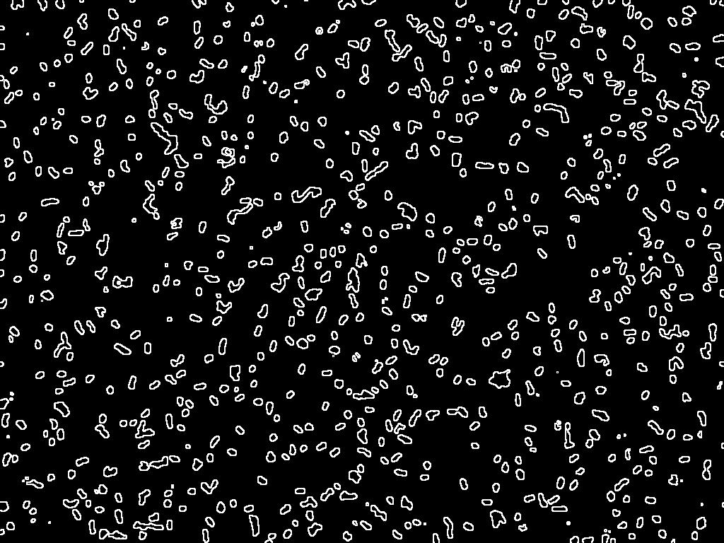
| The image is first changed to black and white, then thresholded and performed with edge detection operation. |
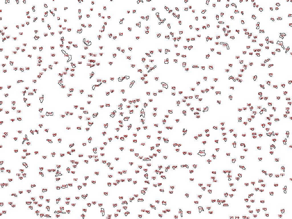
| After performing particle analysis, the particles are outlined and numbered. |
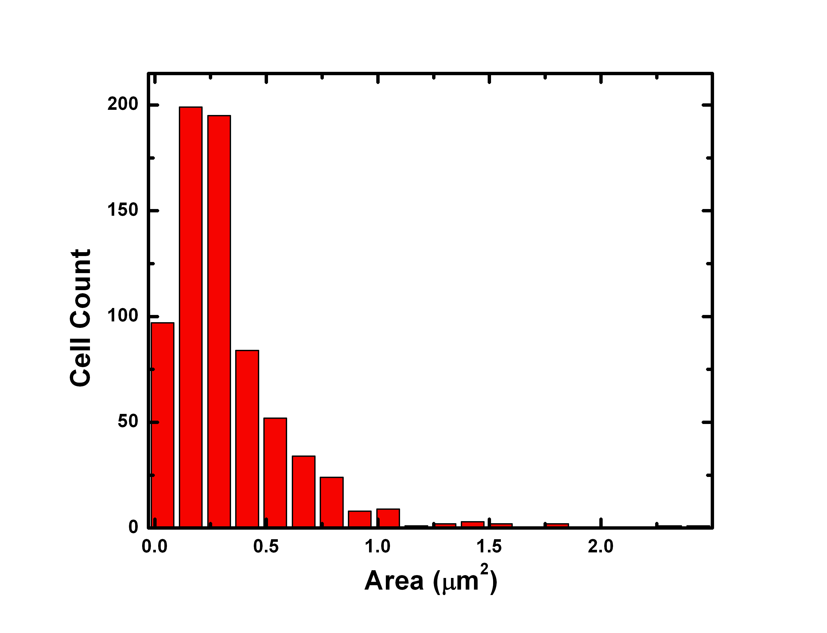
| Plot of particle size distribution. A total of 714 particles are counted with average area of 0.388 ¦Ìm^2. Assuming circular cross section of cells, this converts to 0.7 ¦Ìm diameter. The major error comes from thresholding. Because the cells are not in the same focusing plane, the threshold operation shrink some of the unfocused cells to very small size which reduces the average area of particle. |
{top}
GFP Photobleaching
The green fluorescent protein (GFP) from the jellyfish Aequorea victoria and its mutants have become a very valuable tool for biological studies. A highly visible, efficiently emitting internal fluorophore is generated when appropriate excitation frequency is applied.
Under strong illumination, the fluorophore of GFPs can irreversibly become non-fluorescent. The mechanism involves photoactivated GFP generates endogenous O2, inducing chromophore damage. We studied the GFP photobleaching dynamics by taking a series of images of GFP labeled E. Coli under fluorescent microscope every 10 seconds. During measurement, we tried to adjust focus slightly to compensate for the drifting of focus.
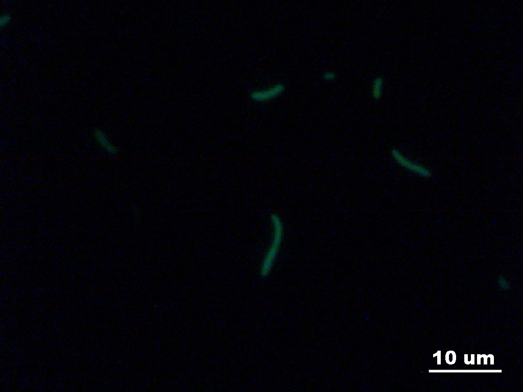
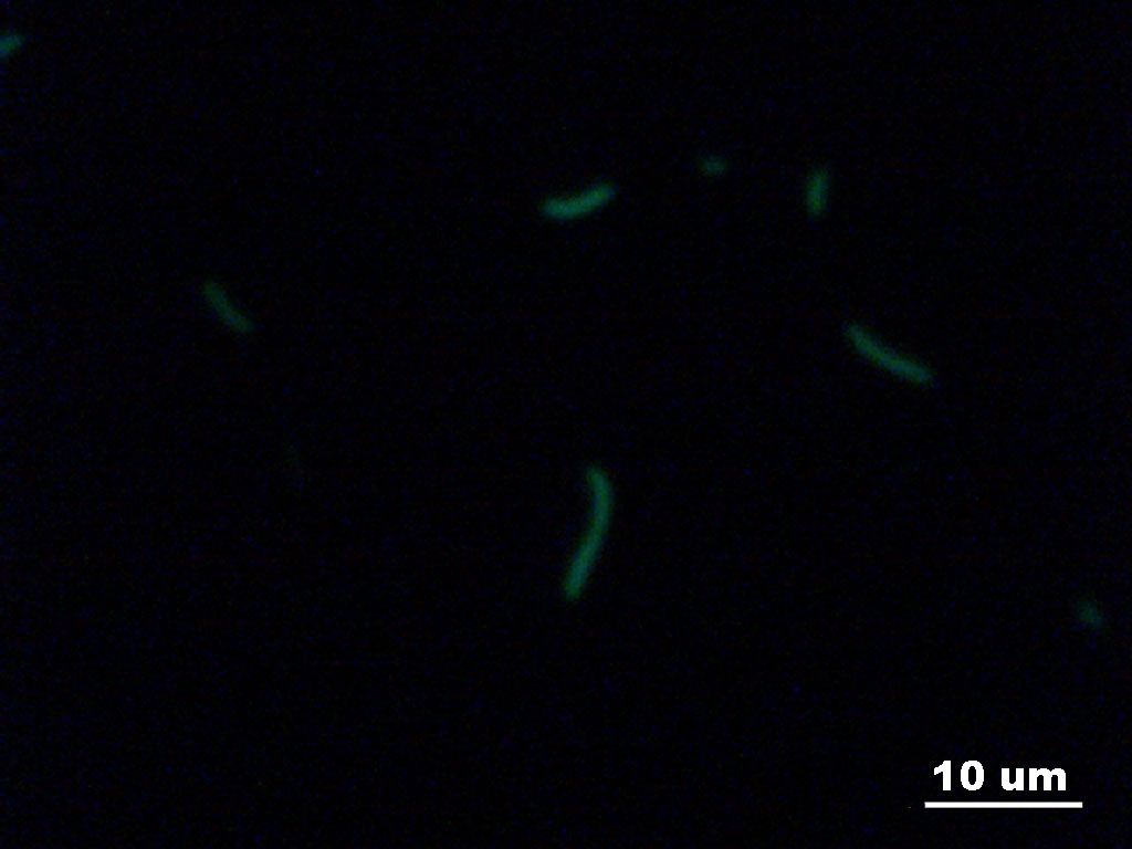
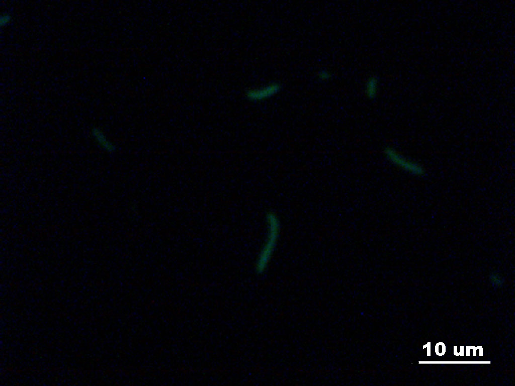
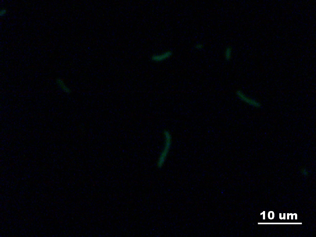
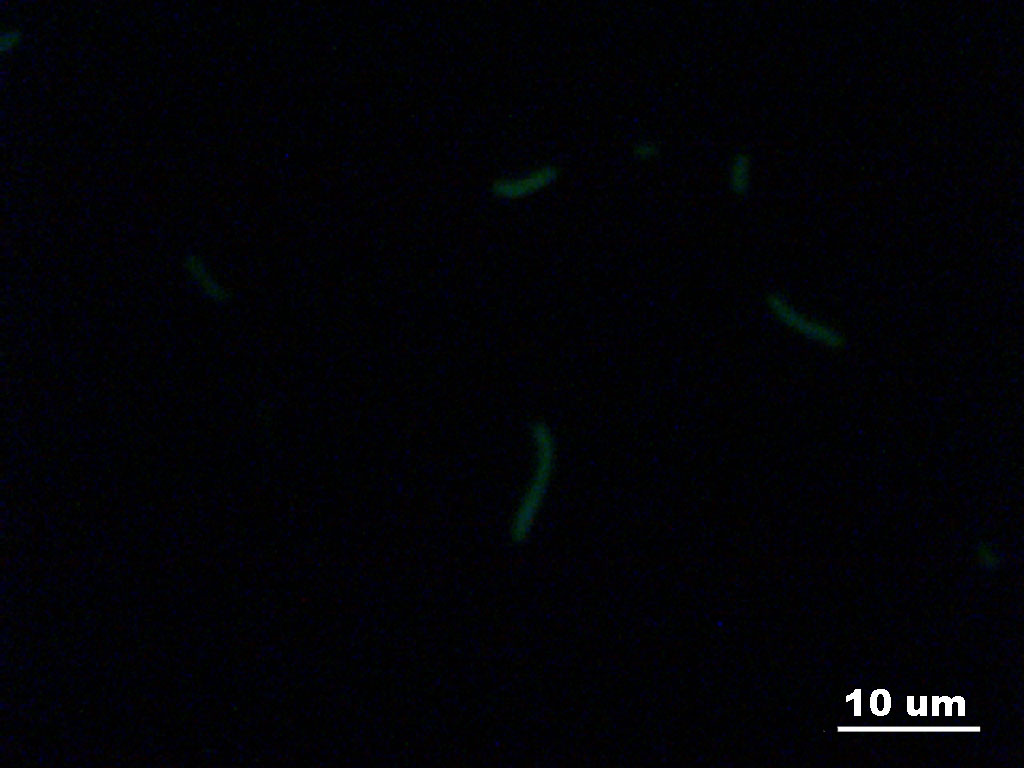
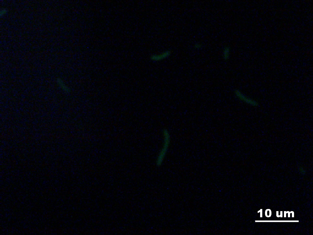
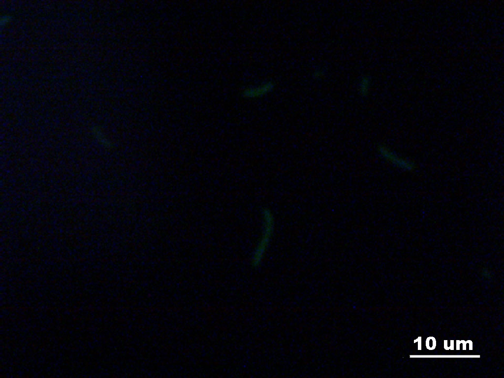
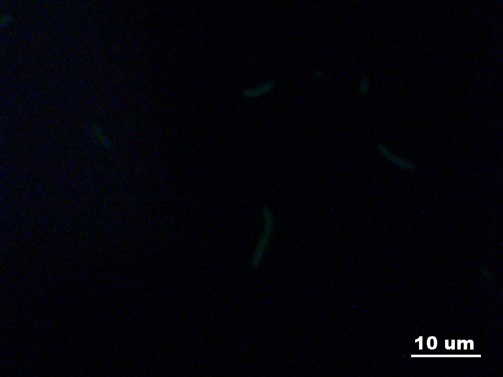
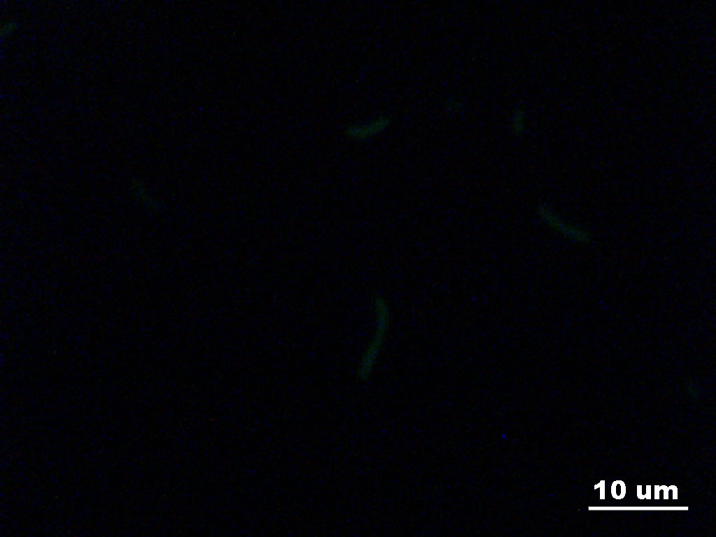
Here is an animated video of stacked images generated by ImageJ. GFP E. Coli movie.
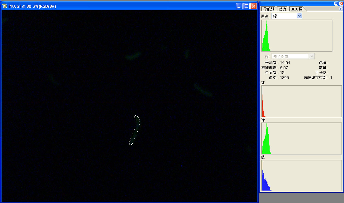
To analyse the image, we use photoshop to select only the areaes of the E. Coli cell at center in all images. We find the green channel histogram of the area and the average pixel intensity.
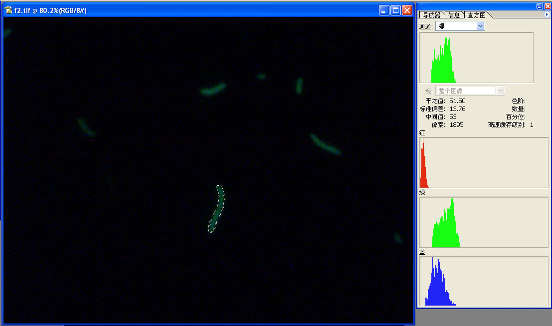
This is the analysis of the first image in photoshop.
This is the analysis of the last image in photoshop.
From the average pixel intensity in selected areas, we plot the light intensity over time. We fit the data to a single mode exponential decay and we calcalculate the time constant of the process to be around 85 seconds. 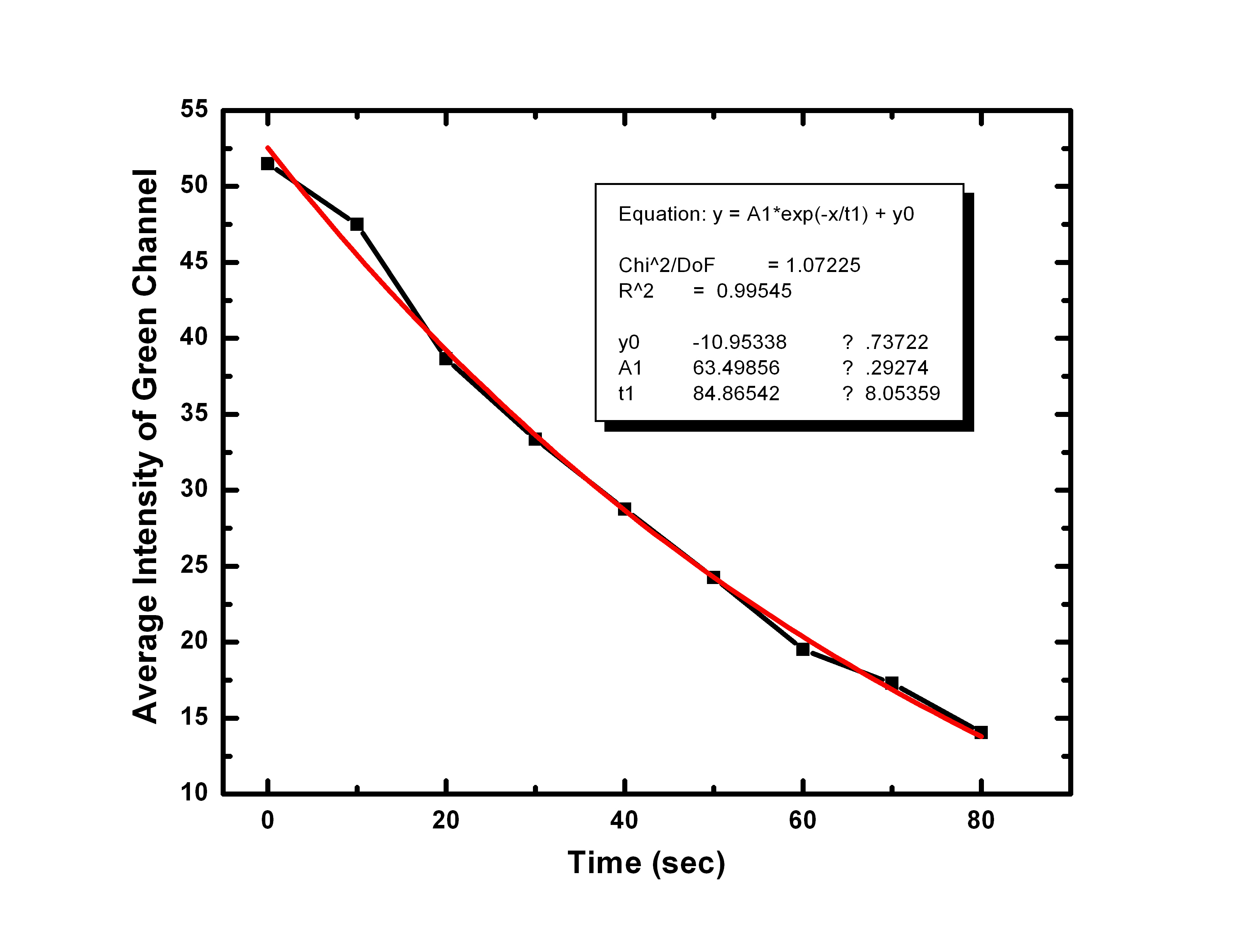
The GFP and its mutants have different time constants. Also the photobleaching dynamics is a function of illumination light intensity. So we should measure the illumination level of the experimental conditions. From a web search on published results, it seems our time constant is at the high end. Normally people report the time constant in seconds and tens of seconds range. It will also be interesting that we could do a sequence search on the GFP we are using and find out if other people have characterize this mutant.
{top}
Fibroblast
A fibroblast is a cell that makes the structural fibers and ground substance of connective tissue. Fibroblasts have a branched cytoplasm surrounding an elliptical, speckled nucleus having 1 or 2 nucleoli. They can can give rise to other cells, such as bone cells, fat cells, and smooth muscle cells. The fact that fibroblasts easily proliferate makes them a popular cell type for cell cultures in biological research. The following pictures were taken using the Zeiss microscope (40x). The actin was stained with FITC, and the mitochondria with Texas Red
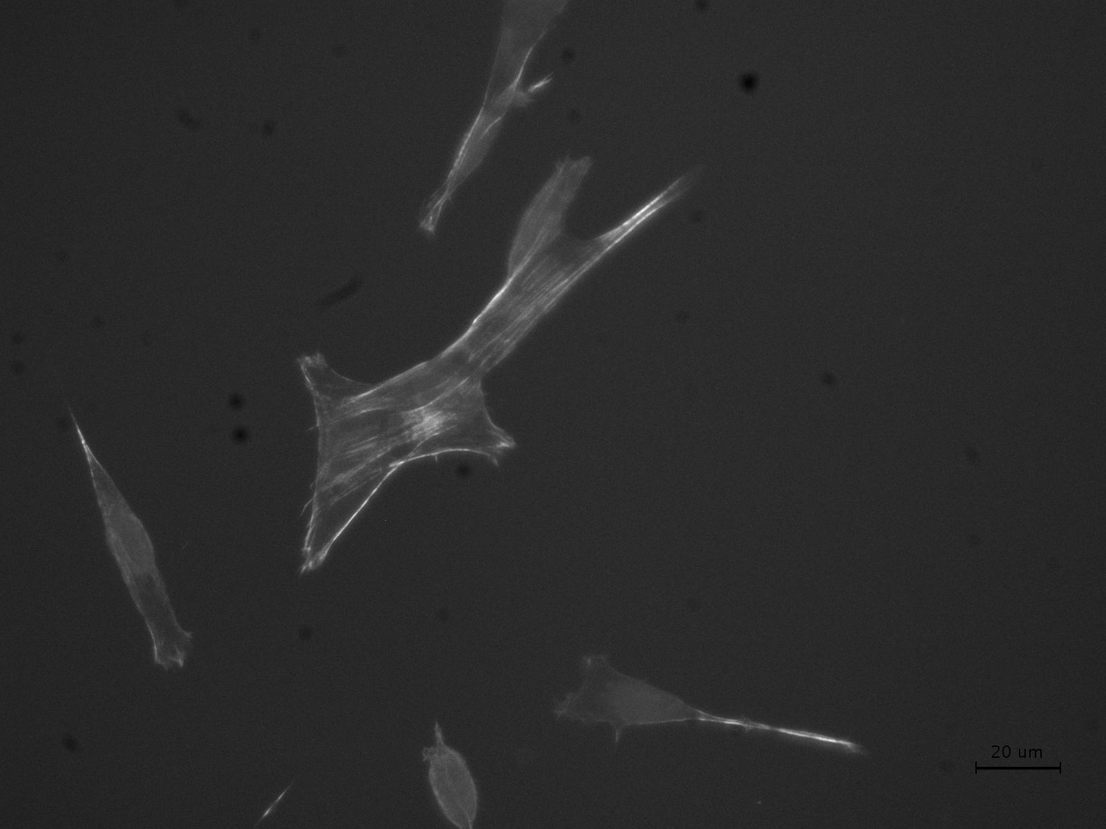
Actin
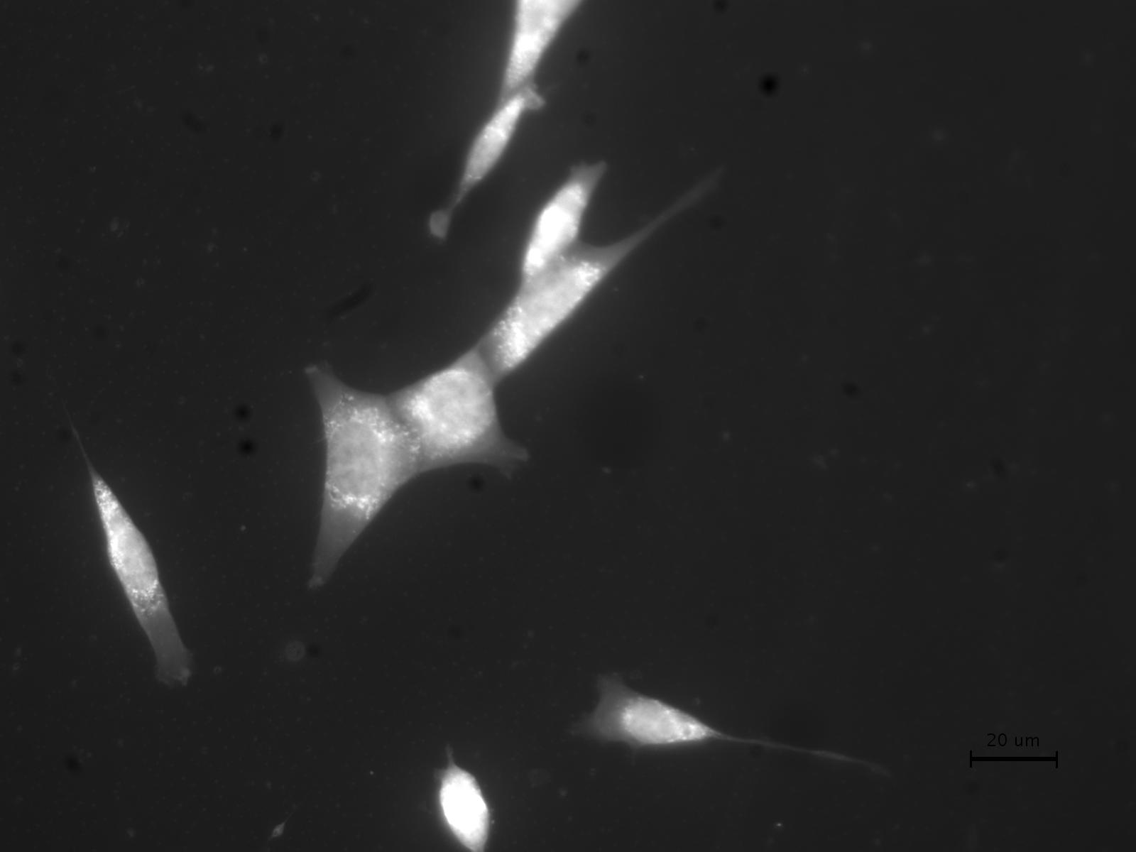
Mitochondria
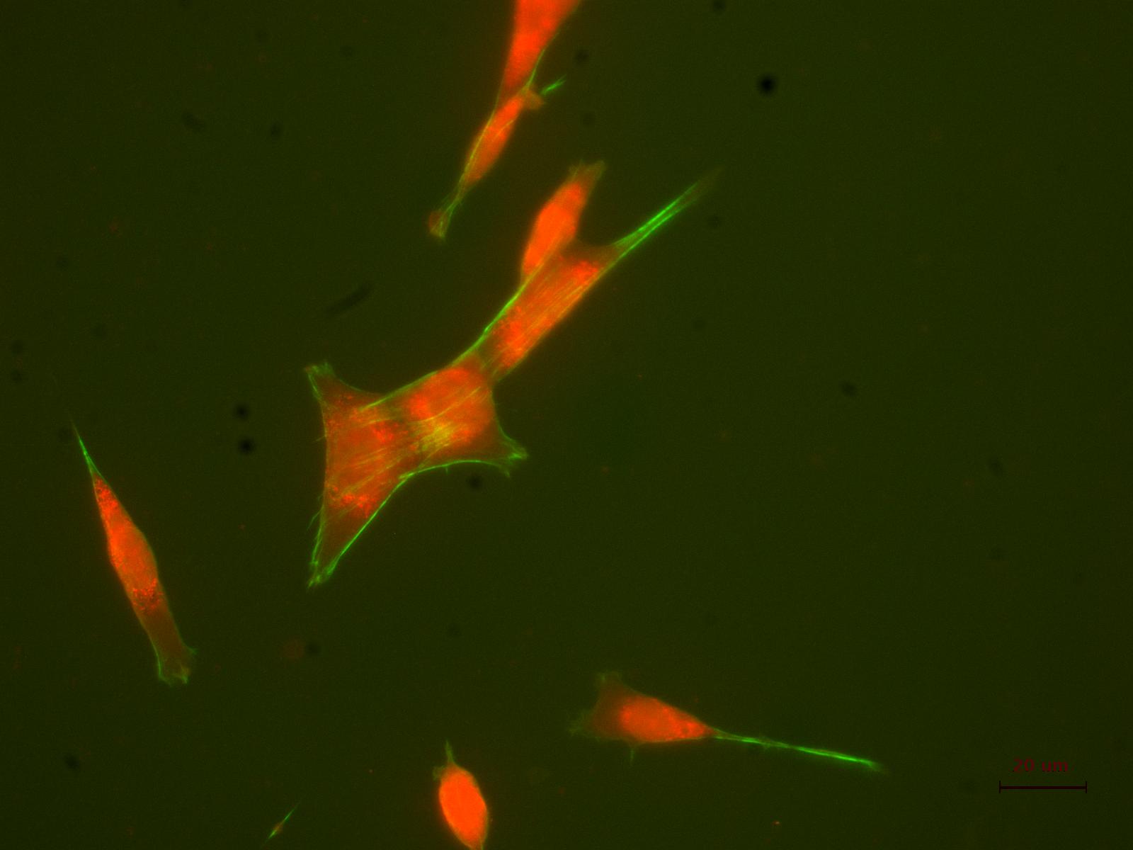
Actin + Mitochondria
With these pictures, we could estimate the length of the fibroblast cells (using our microscope calibration):
Cell |
Length in pixels |
Length in um |
1 |
399.44 |
105 |
2 |
413.90 |
109 |
3 |
297.44 |
78 |
With the following picture we could estimate the diameter of the nuclei:
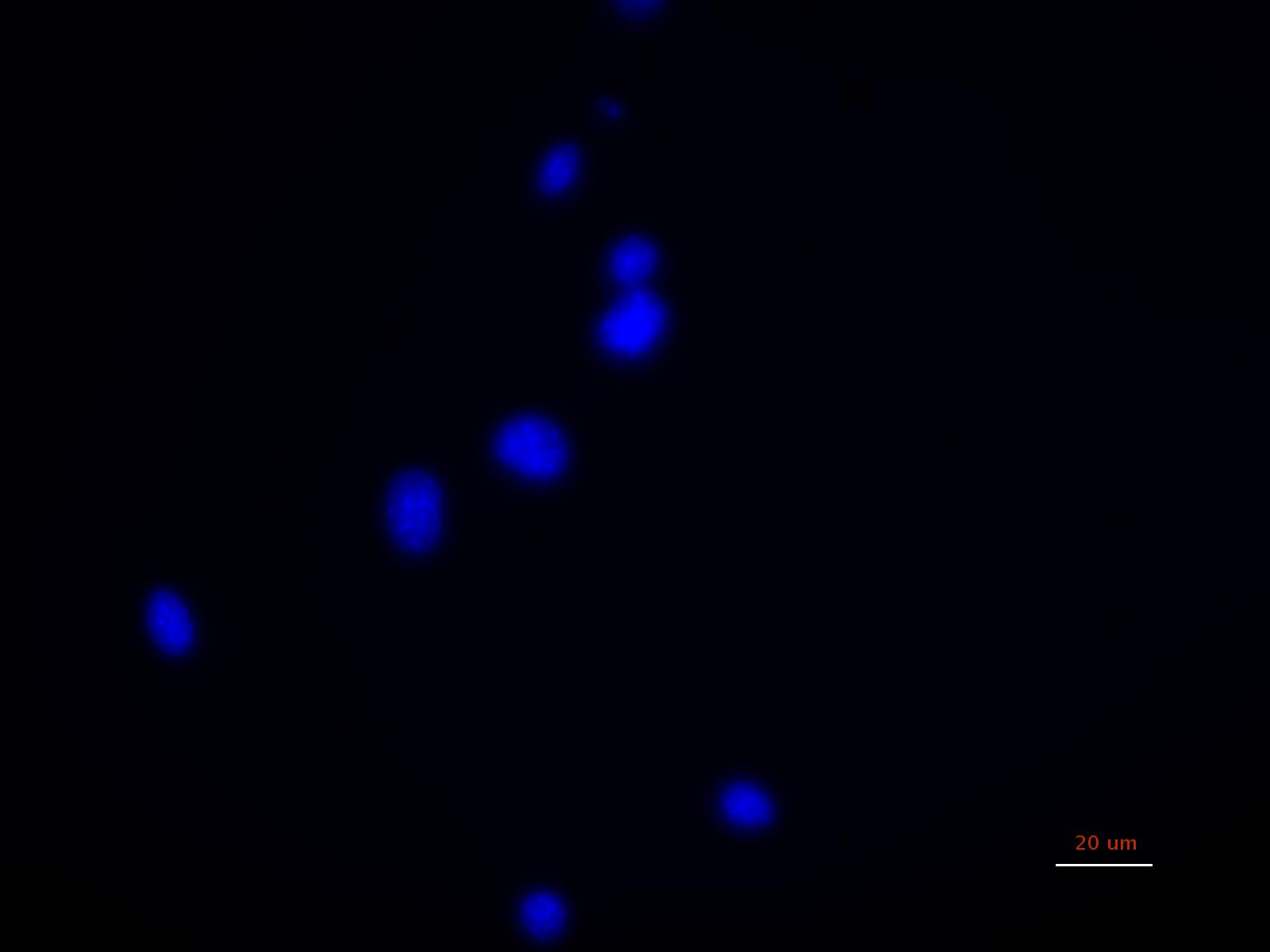
Diameter of nuclei |
Nucleus |
Length in pixels |
Length in um |
1 |
104.69 |
28 |
2 |
83.57 |
22 |
3 |
59.06 |
16 |
4 |
69.43 |
18 |
5 |
89.64 |
24 |
6 |
82.8 |
22 |
7 |
53.85 |
14 |
8 |
74.73 |
20 |
Average |
20.5 |
{top}
Drosophila melanogaster
Drosophila melanogaster (fruit fly) is a little insect about 3mm long. It is one of the most valuable of organisms in biological research, particularly in genetics and developmental biology. Its benefits as a model system include availability of large number of mutants, relative short life cycles, easy maintainess and well known genetics. Its development cycle is 14 days at 25 Celsius degree. The drosophila egg is about half a millimeter long. It takes about one day after fertilisation for the embryo to develop and hatch into a worm-like larva. The larva eats and grows continuously, moulting one day, two days, and four days after hatching (first, second and third instars). After two days as a third instar larva, it moults one more time to form an immobile pupa. Over the next four days, the body is completely remodelled to give the adult winged form, which then hatches from the pupal case and is fertile within about 12 hours.
Apterous is a selector gene which establish the autonomy and direct the development of compartments with respect to other adjoining compartments. Apterous gene has at least two distinct outputs. First, Apterous is responsible for making the dorsal cell distinct from ventral, a property that may be due to its activating the gene Dorsal wing. Second, Apterous regulates the expression of boundary determining proteins such as Fringe and Serrate. Serrate is thought to act as a ligand of Notch. Serrate appears to signal from dorsal to ventral cells to elicit the production of a long range morphogen, perhaps Wingless. Fringe is actually responsible for the induction of Serrate.
Scarlet is a homozygous recessive mutant which prevents the production of brown pigment. The mechanism of 3-hydroxykynureneine storage during larval life is defective, so that this compound is excreted at an abnormally high rate.
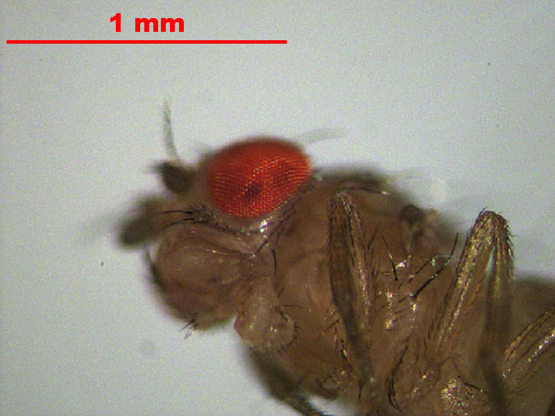
Mutant apterous
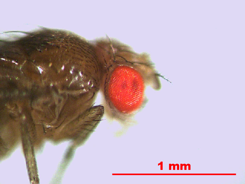
| Mutant scarlet. From this image, we estimate the dimension of the eye is 300 um. |
{top}
Yeast
Yeasts constitute a group of single-celled fungi. There are more than one thousand species of yeasts. The most commonly used yeast is Saccharomyces cerevisae, which is used to produce wine, bread and beer. It is also a model organism, used in genetics and molecular biology studies.
They can reproduce asexually through budding, or sexually. What we were interested in observing in this experiment was their cell cycle. Unfortunately, it was only possible to have a simple movie. Yeast_movie.
{top}