|
Background:
Microscopy is a useful tool commonly used in understanding cell
biology. In this session, techniques such as dark field, bright
field and fluorescent imaging were introduced, and the importance of
scale bars and calibrations was strongly enforced. A feel for the
dimensions and scale of things was also extensively explored.
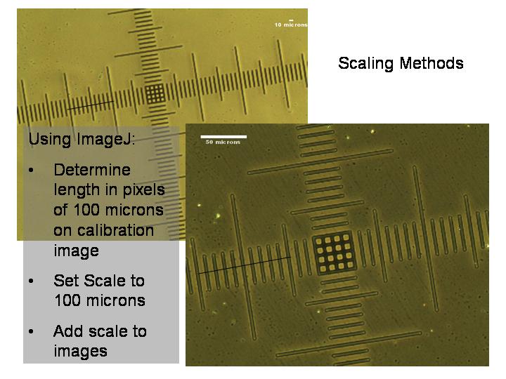
Methods:
1) Calibration of individual microscopes was carried out using a
stage micrometer for different magnification powers (i.e. 10x, 20x,
40x and 100x).
2) Various organisms are placed on glass slides, and covered with
glass coverslips and observed under various magnifications.
3) Using Image J, the pixels on the images can be calibrated to the
actual dimension of the specimen.
Results and Discussion:
Four organisms were studied under various microscopes and magnifications:
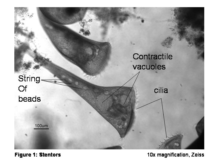
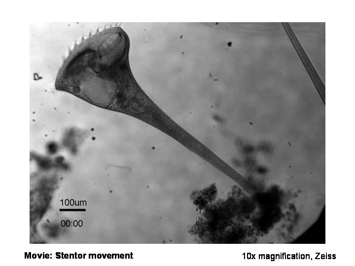
a) Stentor: The largest single-celled protozoa found in water.
Interesting features such as propelling cilia, contractile vacuoles
and macronuclei were quantitatively observed. Dimensions are
characterized in the table below. Images were acquired on a
Zeiss Axio microscope at 10x magnification.
|
|
Average Dimensions |
|
Length of stentor |
1
mm |
|
“Trumpet” region |
350 microns |
|
Contractile vacuoles |
110 microns |
|
Macronucleii (beads on a string) |
33
microns |
Click on figure below for movie (9 MB):
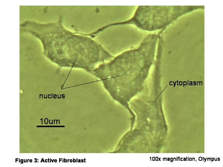
b) Fibroblast: Derived from mesenchymal tissue, fibroblasts are
building blocks for structural fibers and connective tissues.
Features such as cytoplasm, nucleus and cell spreading were
observed. The diameter of a nucleus is about 13 microns and the
average length of a cell ranges from 25-50 microns. Images
were acquired at 100x magnification on an Olympus CX41 microscope
using Moticam 1000 camera and software.
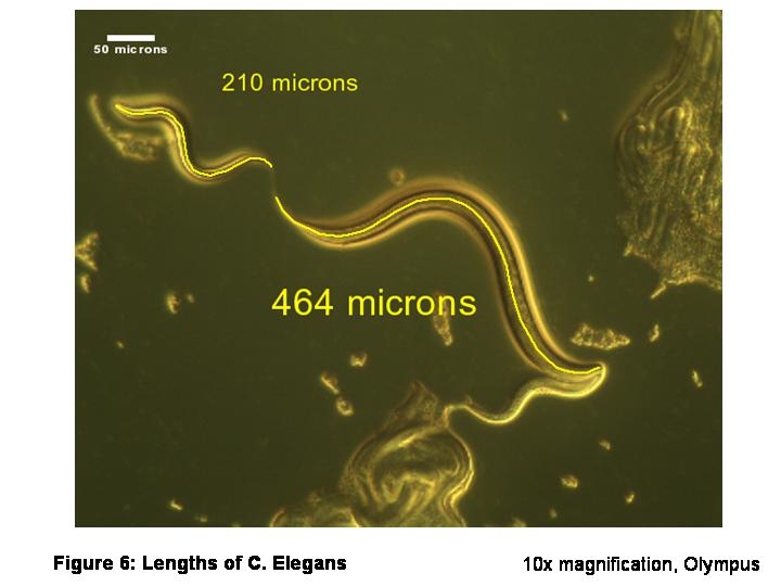
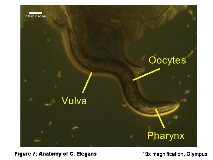
c) C. Elegans: First ever to have its genome sequenced. Features
such as the mouth, pharynx, intestine, gonad, and collagenous
cuticle were observed. Speed of C. elegans movement was also
calculated. The length of these C. Elegans can range from 210 – 464
microns, with the average body length of 79 microns. Images were
acquired at 10x magnification on an Olympus CX41 microscope using
Moticam 1000 camera and software.
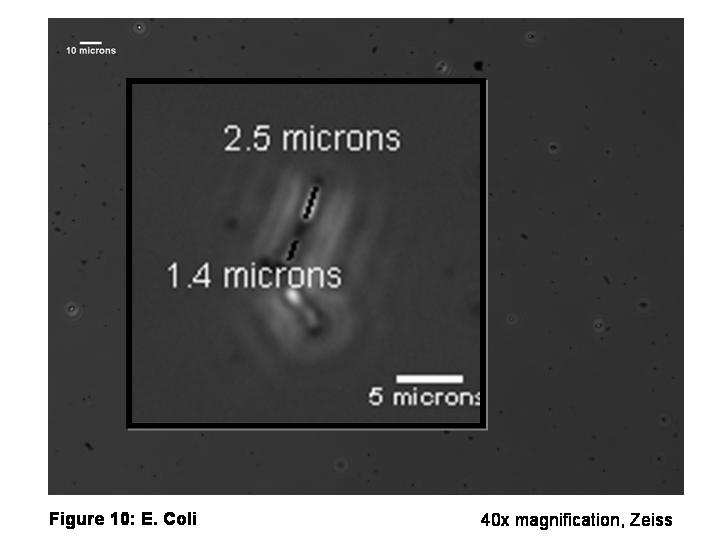
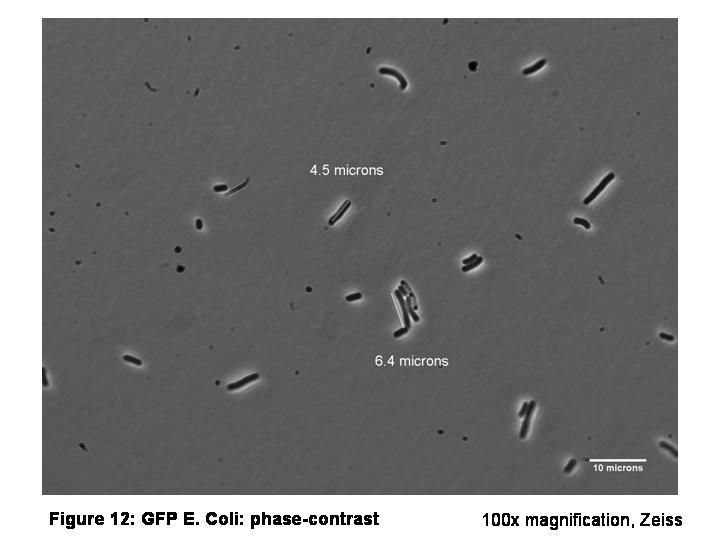
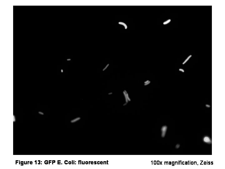
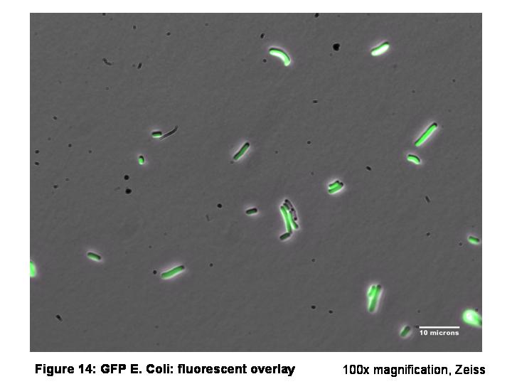
d) E Coli (bacteria): Most commonly studied prokaryote. Stained with
green fluorescent protein (GFP), E coli cells were visualized under
the Zeiss microscope. Photobleaching and the movement of E coli
cells were also observed. The average length of an E coli cell
ranges from 1.4 – 6.7 microns. Images were acquired on an Zeiss Axio
microscope at 40x and 100x magnification.
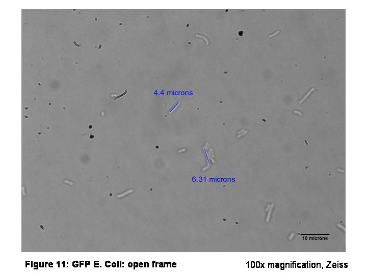
Conclusion:
More samples could be measured to give a more accurate picture of
the average dimensions of each organism. To overcome fast photo
bleaching of the GFP-E coli cells, cells should be located on bright
field mode, before rapidly switching to fluorescent mode to obtain a
picture.
|