|
|
Bioengineering Bootcamp
September 2007
In September 2007 we joined forces with Joel Swanson (University of Michigan), Elaine Bearer (Brown University), Michael Roukes (Caltech), and Scott Fraser (Caltech), and hosted the first bootcamp for the incoming bioengineering class at Caltech as part of their orientation. Joining us were also students from UCLA as well as postdocs from Caltech. This six day crash course was intended to expose bootcampers to both advanced techniques in microscopy and molecular biology as well as cutting edge research projects pursued in our lab and various other labs at Caltech. The bootcampers participated in an advanced Matlab course and learned about particle tracking, creating composite images, curve fitting and more, and also learned about Vector NTI, a useful software tool for designing experiments in molecular biology. We also heard talks by Joel, Elaine and our own Dr. Heun Jin Lee. The days were long but exhilarating, and the course culminated on the sixth day when bootcampers reported the discoveries that they made in their individual projects while wearing their choice of special costume! Here is a quick glance ats what we did.
Day 1: - Microscopy: The
Size of Things. An important step in understanding new
scientific concepts is learning the scales of the problem.
Over what spatial scales do biological processes occur? How
much energy is consumed? In our courses be always begin by
looking at various cells and organisms to discern the overall
size, sizes of organelles, and rates of whole-cell and intracellular
movement, using a variety of light and fluorescence microscopy
techniques.
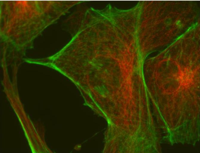
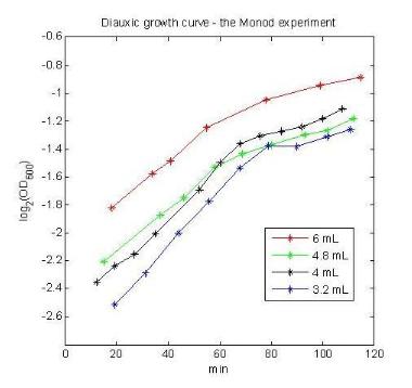
- Microscopy: The Rate of Things. Just like in The Size of Things, here we try to get the students acquainted to the time scales of biological processes. We looked at some pond scum (video below), Drosophila melanogaster, C. elegans, Dictyostelium, and bacteria and yeast. - Spectrometry: Examining the rate of things. We can infer much about the rate of biological processes not just by directly observing them through a microscope, but also through less direct measurements, such as monitoring the optical density of a population of growing cells. Much can be deduced about the physiology of bacterial cells just by monitoring how population density changes over time. In this part of the bootcamp we set to recreate one of the most famous experiments in biology conducted by Jaques Monod that suggested the notion that bacterial cells do not waste resources to make proteins if they are not needed. The idea in this experiment is to place E. coli cells in a medium where the organic source is the limiting factor and is composed of a mixture of two carbohydrates, such as glucose and lactose. E. coli will first utilize the preferred energy source (glucose in this case) while suppressing proteins required utilization of the second sugar. Once glucose is exhausted form the medium the cell will undergo a lag period where it needs to synthesize the proteins required for intake and digestion of the second carbon source (lactose). Once these enzymes are produced exponential growth is resumed and the cells continue to double, albeit at a lower doubling rate. In the figure shown the bootcampers demonstrate the diauxic shift for various inoculation volumes of starter E. coli cultures growing in a glucose/lactose medium.
Day 2: - DNA
Science. Molecular biology has progressed at an amazing
rate in the last two decades yeilding a set of tools that
allow us to manipulate DNA in a very controlled way. The
aim of this section of the courses is to show a set of examples
of the different tools that can be used to solve a wide variety
of problems. Our claim is that, at least when dealing with
E. coli, it is mostly about asking the right question rather
than developing new techniques.
- Quantitative PCR . To analyze the PCR amplification of the target gene in a more accurate way some of the bootcampers that were familiar with conventional PCR monitored this reaction by means of Quantitative PCR or QPCR for short. Here a fluorescent stain for dsDNA is added to the reaction and enables us to monitor the number of replicates as the cycle progresses. The figure above shows the amplification plots of the targets (early) and no targnet controls (late).
|
|||
|
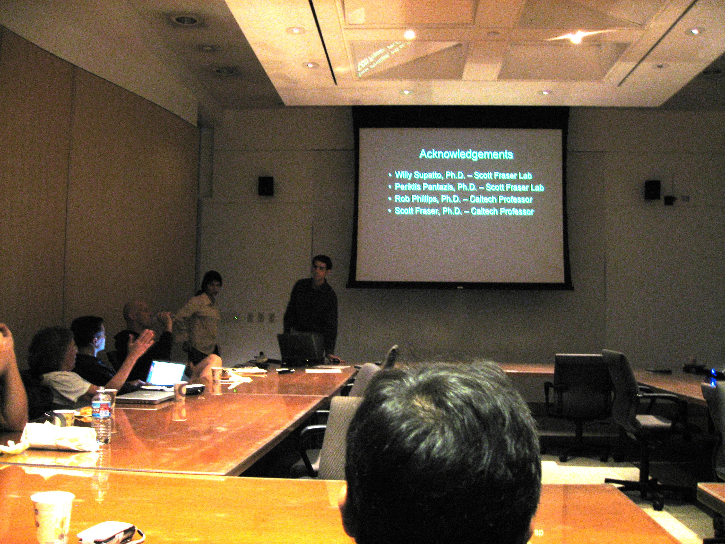
Days 3, 4, and 5: - Advanced Projects. The students split into smaller groups to carry out a variety of very interesting projects. The projects that the students worked on are listed below. For more details on each of the projects, click on each link. Macrophages Joel Swanson, Heun Jin Lee, Dr. Barak Dayan (PMA), Alborz Mahdavi (BE), Jonathan Sternberg (BE) Drosophila FRAP Scott Fraser, Laki Pantazis, Willy Suppatto, William Dempsey (BE), Shima Hajimirza (BE) Tethered Particle Motion Steph Johnson (BMB), Geoffrey Lovely (BMB), David Schwab (Ph), Sung Wook Woo (BE) Limulus sperm dynamics Elaine Bearer, Drew Kennedy (BE), Niema Pahlevan (BE) Single cell calorimetry Michael Roukes, Wonhee Lee, Nakul Reddy (BE) Microfluidics embedded biosensors Michael Roukes, Jessica Arlett, Indira Wu (BE), Zhaoyan Zhu (Ch) Cilia reconstitution Dave Wu (BE), Andrew Fung, Hsiang-Wei Lu Day 6: The last day every group gave a presentation showing everybody what they had accomplished during this intense week.
|
| Copyright Phillips Group 2005-2008 |