.: Introduction
The goal of this lab was to visualize mitosis
in vivo by employing time-lapse microscopy of the fertilization and
subsequent nuclear division of the embryo using a high resolution camera.
The following types of sea urchins were utilized in the experiments:
Lytechinus variegatus
(green sea urchin)
Lytechinus variegatus are handsomely pink and white, with an explosion of
short spines emanating from a fat round body. Eggs are remarkably clear and
easy to study, particularly for demonstrating mitotic spindles. Huge volumes
of eggs can be produced from a single large specimen, hence it is also
prized by seafood loving gourmets. Specimens are fertile May through
September. |
 |
Arbacia punctulata (purple-spined
sea urchin)
This urchin bristles long, sharp, formidable
looking spines as it rapidly moves about the aquarium. Their powerful teeth
scrape away algae, and chew into sponges. Although they add action and
beauty to the salt water aquarium, they have been classically used in
embryology. Specimens are fertile mid-January through April. |
 |
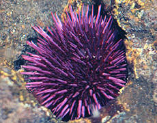
Strongylocentrotus purpuratus (purple sea urchin)
Purple sea urchins are found on the
pacific coastline from Alaska to Cedros Island, Mexico. The purple sea
urchin thrives amid strong wave action and areas with churning aerated
water. They must be kept in cold conditions. This urchin has adapted
the ability to burrow itself into the substrate, often times rock. It uses
its five bony teeth in concert with its spines to slowly gauge and scrape
away at the substrate. The result is a depression in the substrate into
which the rest of the urchin can settle with a firm hold. This is a unique
feature that can sometimes prove deadly. When S. purpuratus is young, it may
begin to scrape into the substrate. As it grows, the urchin may find that it
has trapped itself for life. January, February, and March are the primary
reproductive months. |
 |
.: Methods & Results
Slide Preparation and Rehydration
Based on studies from Week 1 & 2, one of the
biggest issues we faced with time-lapse microscopy was dehydration of the medium
that the cells were in due to heat and light from the microscopy, both necessary
to visualize the processes. To prevent dehydration we employed two
suggestions:
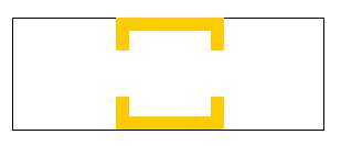
1. Provide a slide well using molten wax to
increase the volume of artificial sea water between the slide cover and the
slide as shown below:
|
 |
2. Then monitor the slide every 30 minutes to
determine the degree of dehydration. If the slide begins to dehydratre,
use a very small pipette tip and draw out approximately 10 uL of artificial
sea water and slow push it under the slide. Let diffusion even out the
dried portions of the slide. Be careful not to expel the sea water too
quickly on to the slide otherwise the image will shift due to the force of
the flow. |
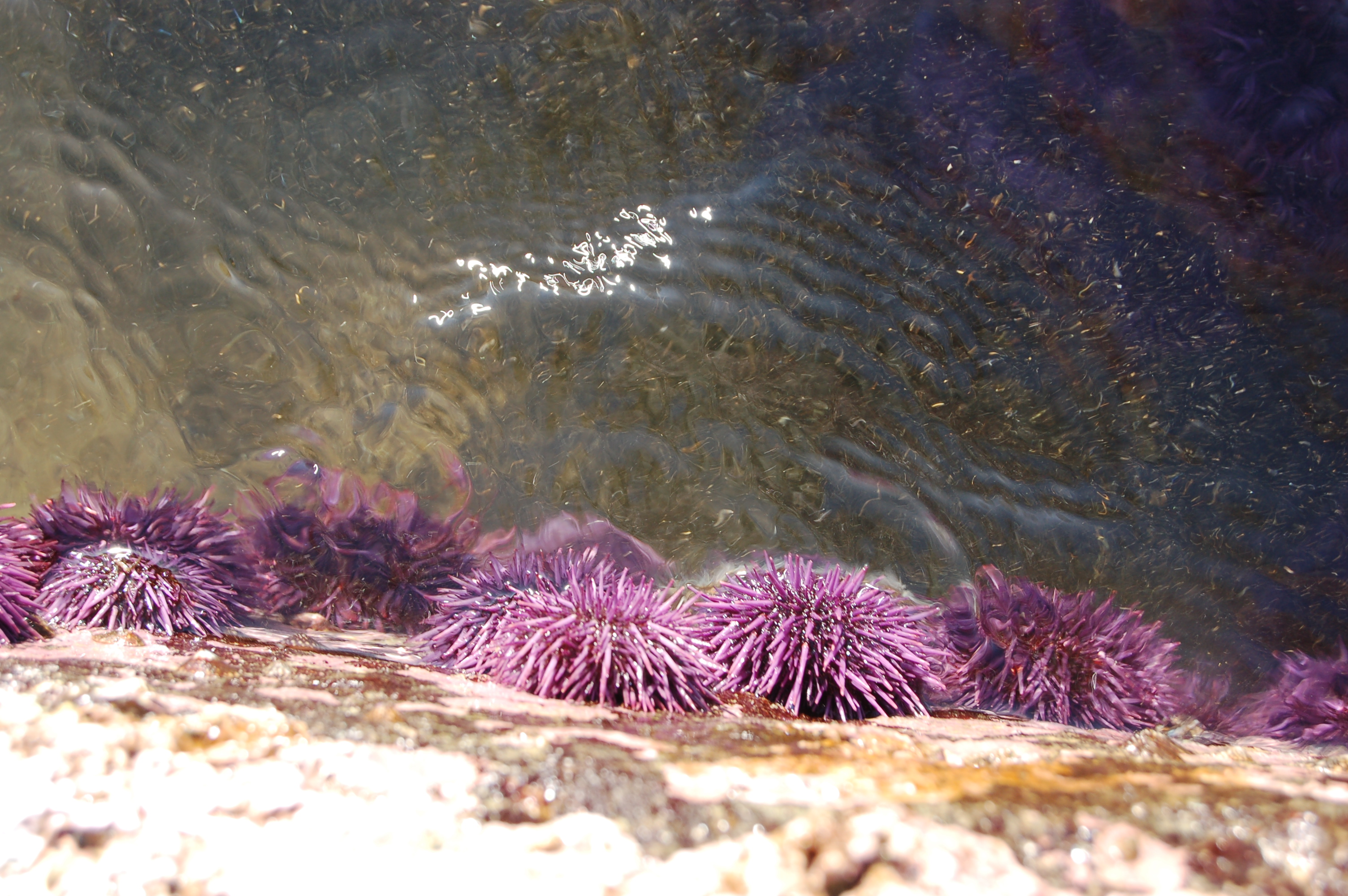
Strongylocentrotus purpuratus (purple sea urchin)
Efforts were made
to obtain purple sea urchin as they are abundant along the California coastline
and they are in the ideal time of fertility. A quick road trip was made to
the Palos Verdes Peninsula, just south of San Pedro. At the tip of the
peninsula,
White Point Beach is home to some of the most diverse tidepools on the West
Coast. Hundreds of purple sea urchins could be found there (see
Picture Gallery). It is important to arrive at
the tide pools at low tide (check
tide tables!) Approximately 40 sea urchins were collected, transported
at sea water temperature, and transferred to the Keck 040 urchin tanks.
Unfortunately there was no way to control the temperature in the tanks.
All of the sea urchins perished before they could be utilized for
experimentation. Furthermore, fertilization and embryology would have to
occur under cold water conditions, making this a difficult
Arbacia punctulata (purple-spined
sea urchin)
Although the the purple-spined sea
urchin were vey hearty, fertilizing them proved to be very difficult.
Several attempts were made to fertilize the species but all attempts were either
unsuccessful or abortive. Either the eggs would not fertilize at all or
they would begin mitosis but would stop at some time in the process. In
the movie shown below, on the left, you can see that the cell begins to go
through telophase but at some point it no longer proceeds and eventually fuses
back into one cell again. A similar deformity occurs in the movie below,
right, where during telophase, the process is "corrupted" as can be seen from
the shape of the inner membrane of the cell.
![]()
![]()
Deform.avi (2.7MB)
Deform2.avi (4.3 MB)
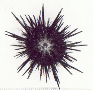
Lytechinus variegatus
(green sea urchin)
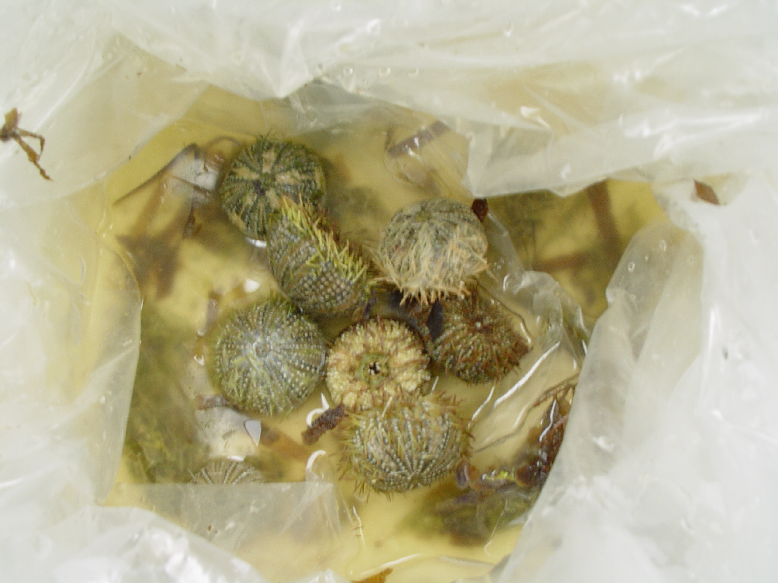 |
Fortunately, the green sea urchin was much
more amenable to fertilization. The only difficulty with this part of
the study was that 90% of the sea urchins shipped from Carolina Bio were
dead on arrival (see image to the left). Only two of the twenty sea
urchins survived the journey of which we were very fortunate to have one
female and one male. The fertilization was successful and the images
provide an excellent view of mitosis. The nucleus can be identified
and followed. Unfortunately at the very end of the images, dehydration
does take place but not before exceeding well over 6 cell cycles.
|
![]()
Success.avi (9MB)
Web site contents © Copyright AEK 2006, All rights reserved.
Website templates
|